Preview
Hüseyin Akbulut, MSc (2026). The Molecular Mechanism of Muscle Contraction. Sporeus. Retrieved, November 17, 2026. https://sporeus.com/en/sport/molecular-mechanism-of-muscle-contraction/
Skeletal muscle contraction is the basic physiological process behind human movement. Every stride, every jump and every lift depends on the coordinated work of thousands of molecular motors inside the muscle fibres.
Table of Contents
The Sliding Filament Theory
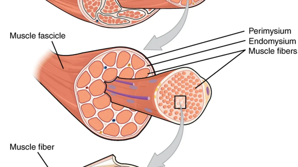
Proposed in 1954 by Huxley and Hanson, the sliding filament theory remains the most widely accepted explanation of muscle contraction. On this account, contraction occurs as the thin filaments (actin) slide over the thick filaments (myosin). Sarcomere length shortens, but filament lengths do not change. Each myofibril within a muscle fibre is made up of thousands of sarcomeres arranged end to end. The sarcomere is the functional unit between one Z line and the next. During contraction the A band stays constant while the I band narrows and the H zone disappears.
The Cross-Bridge Cycle
The cross-bridge cycle has four main steps. In attachment, the myosin head binds to its site on actin, which becomes possible when calcium binds to troponin C. In the power stroke, the myosin head changes conformation and pulls the actin filament; ADP and inorganic phosphate are released. In detachment, a new ATP binds to myosin and myosin releases actin. In the recovery step, ATP is hydrolysed and the myosin head returns to its cocked position.
- Energy cost: each cycle consumes one ATP and produces roughly 10 nm of sliding.
- Rate: the cycle can repeat about five times per second.
- Myosin ATPase: the key enzyme setting contraction speed; its activity varies with fibre type.
- Rigor mortis: in the absence of ATP, myosin cannot detach from actin.
Calcium Signalling and Excitation–Contraction Coupling
An action potential arriving from the motor neuron triggers acetylcholine release at the neuromuscular junction. The action potential in the muscle fibre membrane spreads deep into the cell through the T-tubules. Voltage sensors in the T-tubules activate ryanodine receptors in the sarcoplasmic reticulum, and a large amount of Ca²⁺ is released into the cytoplasm. At rest, cytoplasmic Ca²⁺ is about 10⁻⁷ M; during active contraction it rises to 10⁻⁵ M. Calcium binds to troponin C, tropomyosin shifts, and the myosin binding sites are exposed. As contraction ends, the SERCA pump transports Ca²⁺ back into the sarcoplasmic reticulum.
The Roles of ATP and the Fibre Types
| Role | Mechanism | Without ATP |
|---|---|---|
| Cross-bridge cycling | Myosin ATPase hydrolysis | Rigor mortis |
| Ca²⁺ reuptake | SERCA pump | Sustained contraction |
| Na⁺/K⁺ pump | Membrane potential | Loss of excitability |
In intense exercise the rate of ATP turnover can rise to 100 times the resting value. The creatine phosphate system, anaerobic glycolysis and oxidative phosphorylation meet that demand. Type I fibres resist fatigue thanks to their low ATPase activity, while Type II fibres contract quickly but fatigue fast. Endurance training accelerates the shift from IIx towards IIa, and strength training increases myosin heavy chain synthesis. These adaptations begin within weeks, but the full effect takes months. Understanding the molecular basis of muscle contraction matters in every area from training design to injury management.
References
- Huxley, H. E. (1969). The mechanism of muscular contraction. Science.
- Huxley, A. F., & Niedergerke, R. (1954). Structural changes in muscle during contraction. Nature.
- Gordon, A. M., Huxley, A. F., & Julian, F. J. (1966). The variation in isometric tension with sarcomere length in vertebrate muscle fibres. The Journal of Physiology.
- Lieber, R. L. (2002). Skeletal Muscle Structure, Function, and Plasticity. Lippincott Williams & Wilkins.

Hüseyin Akbulut is the founder of Sporeus and author of THRESHOLD (EŞİK), a 540-page Turkish-language book on endurance science.
- 540pp THRESHOLD Book
- MSc Sport Sciences
- Marmara University
The Sliding Filament Theory
Proposed in 1954 by Huxley and Hanson, the sliding filament theory remains the most widely accepted explanation of muscle contraction. On this account, contraction occurs as the thin filaments (actin) slide over the thick filaments (myosin). Sarcomere length shortens, but filament lengths do not change.…
The Cross-Bridge Cycle
The cross-bridge cycle has four main steps. In attachment, the myosin head binds to its site on actin, which becomes possible when calcium binds to troponin C. In the power stroke, the myosin head changes conformation and pulls the actin filament; ADP and inorganic phosphate…
Calcium Signalling and Excitation–Contraction Coupling
An action potential arriving from the motor neuron triggers acetylcholine release at the neuromuscular junction. The action potential in the muscle fibre membrane spreads deep into the cell through the T-tubules. Voltage sensors in the T-tubules activate ryanodine receptors in the sarcoplasmic reticulum, and a…