Preview
Hüseyin Akbulut, MSc (2026). ACL Injury Biomechanics — Why It Tears and How to Protect It. Sporeus. Retrieved, October 2, 2026. https://sporeus.com/en/biomechanics/acl-injury-biomechanics-prevention-football/
Introduction
Anterior cruciate ligament rupture is the injury that ends seasons and careers. At 8–12 months of recovery minimum, an ACL tear removes a player from the squad for essentially an entire competitive year. Yet 70% of ACL injuries in football occur without any contact — no collision, no tackle, just a player planting their foot, changing direction, or landing from a jump, and feeling the knee give way. Understanding exactly what biomechanical forces tear the ACL, and why certain movement patterns generate those forces, is the basis for every evidence-supported prevention programme.
Table of Contents
The Science
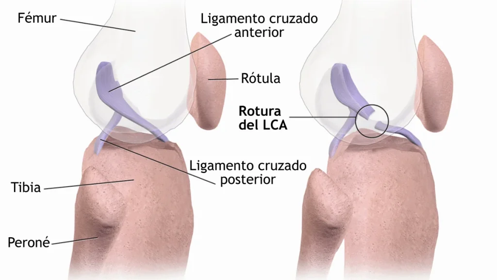
The anterior cruciate ligament is a 3–4 cm intraarticular ligament that connects the femur to the tibia, running anteriorly and medially. Its primary mechanical function: resisting anterior tibial translation relative to the femur (the tibia sliding forward), resisting tibial internal rotation, and contributing to valgus stability. Rupture occurs when one or more of these forces exceed the ligament’s ultimate tensile strength (approximately 2,160 N in adults — though this varies significantly with age and training history).
The ACL injury mechanism: Over 70% of ACL tears in football are non-contact and occur within a narrow biomechanical pattern:
- Knee near full extension (15–40° flexion) at the moment of injury — the quadriceps are powerfully active, pulling the tibia anteriorly (anterior tibial shear force) while the ACL is maximally loaded
- Knee valgus collapse (knee caving inward) — the most consistently observed ACL injury position on video analysis. Valgus creates combined anterior shear and tibial internal rotation — the two forces the ACL resists simultaneously
- Planted foot with hip internal rotation and adduction — typically during a cutting manoeuvre, side-step, or single-leg landing
- Low knee flexion at initial contact during landings — stiff landings without adequate hip and knee flexion shift load to the ACL rather than distributing it through the musculature
Why female players tear ACL 2–6× more often: Three primary factors:
- Q-angle (angle from ASIS to patellar tendon) is larger in females (15–20° vs. 10–15° in males), directing patellar tendon force more medially and predisposing to valgus collapse
- Intercondylar notch width is narrower relative to ligament CSA in females — the ACL may be subject to bony impingement during dynamic loading
- Hormonal effects on ligament laxity — oestrogen receptors in ACL tissue increase laxity during high-oestrogen cycle phases
What Research Says
Hewett et al. (2005) published the landmark prospective ACL prediction study in American Journal of Sports Medicine, using 3D motion analysis during drop-jump testing to screen 205 female athletes before their season. Nine subsequent ACL injuries occurred — and all nine were in players who had shown significantly greater knee abduction moments (valgus loading) at screening. The ability to predict individual ACL injury risk from biomechanical screening was confirmed.
Dempsey et al. (2007) conducted systematic video analysis of ACL injuries across multiple football codes in the British Journal of Sports Medicine, characterising the common injury position as: trunk upright or in slight lateral flexion toward the planted leg, hip internally rotated and adducted, knee near full extension, valgus moment at time of contact. This biomechanical profile forms the basis of targeted movement screening protocols.
Alentorn-Geli et al. (2009) published a comprehensive review confirming the efficacy of neuromuscular training programmes (FIFA 11+, PEP programme) in reducing non-contact ACL injuries by 50–88% in female athlete populations — with the common mechanism of efficacy being improved landing mechanics, reduced knee valgus, and greater knee flexion depth at initial contact.
Did You Know? High-speed video analysis of actual non-contact ACL ruptures (rare footage captures) consistently shows the injury occurring in the first 50ms after foot contact — before any protective muscular activation can occur in response to the stimulus. Muscles contract in approximately 80–120ms in response to a surprise stimulus. This means the ACL can only be protected by pre-contact motor patterns programmed before the ground is reached — not by reactive muscular response. This is exactly what neuromuscular training reprograms: the automated movement pattern before contact.
Applied to Football
ACL biomechanics-informed prevention and movement training:
- Train landing mechanics explicitly. Teach and reinforce soft landings: knee flexion >45° at initial contact, knees tracking over toes, hips loaded rather than stiff knee-dominant landing. Perform this in every warm-up for youth and female players.
- Develop gluteal strength and hip external rotation. Valgus collapse is fundamentally a hip control failure. Glute bridges, clamshells, Copenhagen adductor work, and single-leg squats develop the hip musculature that prevents valgus during cutting and landing.
- Screen with overhead squat and single-leg squat assessment. Knee valgus in single-leg squat is a practical proxy for landing mechanics risk. All players showing valgus collapse in screening should receive targeted correction before high-risk activity.
- Programme lateral and cutting movement mechanics. Cutting mechanics can be trained to produce lower valgus moments — specifically, planting with more knee flexion and taking shorter approach strides before direction change. This is teachable, and the reduced injury rates from neuromuscular programmes prove it works.
- Plyometric volume progression for returning players. Post-ACL reconstruction players returning to high-speed movement have rehabilitated the structural ligament but may not have restored the neuromuscular patterns that protect it. Progressive cutting, landing, and reactive plyometric sequences in the final rehabilitation phase are not optional.
Key Takeaways
- 70%+ of ACL tears are non-contact; they occur at low knee flexion, high valgus, and planted foot position
- The injury mechanism combines anterior tibial shear + tibial internal rotation — the two forces the ACL resists
- Neuromuscular protection must be pre-programmed — reactive muscle response is too slow to protect the ACL
- Female players have 2–6× higher ACL rates; Q-angle, notch width, and hormonal laxity contribute
- Neuromuscular programmes (FIFA 11+) reduce non-contact ACL injuries by 50–88% in compliant groups
References
- Hewett, T. E., Myer, G. D., Ford, K. R., Heidt, R. S., Colosimo, A. J., McLean, S. G., … & Succop, P. (2005). Biomechanical measures of neuromuscular control and valgus loading of the knee predict anterior cruciate ligament injury risk. American Journal of Sports Medicine, 33(4), 492–501.
- Dempsey, A. R., Lloyd, D. G., Elliott, B. C., Steele, J. R., & Munro, B. J. (2009). Changing sidestep cutting technique reduces knee valgus loading. American Journal of Sports Medicine, 37(11), 2194–2200.
- Alentorn-Geli, E., Myer, G. D., Silvers, H. J., Samitier, G., Romero, D., Lázaro-Haro, C., & Cugat, R. (2009). Prevention of non-contact anterior cruciate ligament injuries in soccer players: a systematic review. Knee Surgery, Sports Traumatology, Arthroscopy, 17(8), 859–879.
—
Next in Series: Article 54 — Energy Availability and RED-S — When Footballers Don’t Eat Enough
Introduction
Anterior cruciate ligament rupture is the injury that ends seasons and careers. At 8–12 months of recovery minimum, an ACL tear removes a player from the squad for essentially an entire competitive year. Yet 70% of ACL injuries in football occur without any contact —…
The Science
The anterior cruciate ligament is a 3–4 cm intraarticular ligament that connects the femur to the tibia, running anteriorly and medially. Its primary mechanical function: resisting anterior tibial translation relative to the femur (the tibia sliding forward), resisting tibial internal rotation, and contributing to valgus…
What Research Says
Hewett et al. (2005) published the landmark prospective ACL prediction study in American Journal of Sports Medicine, using 3D motion analysis during drop-jump testing to screen 205 female athletes before their season. Nine subsequent ACL injuries occurred — and all nine were in players who…
Applied to Football
ACL biomechanics-informed prevention and movement training: